Craig writes:
Greetings Twiv Team,
I’m a long time listener (all 491 episodes) and huge fan of the show. I’m writing from St. Louis where it’s a beautiful spring day: 78F with clear blue skies, winds from the SE at 15 mph, 60% humidity and a dew point of 62 degrees. Sadly though, the cherry blossoms have faded.
I wanted to follow up on TWiV 491 where you kindly covered our paper on norovirus infection of tuft cells. Given space constraints we weren’t able to elaborate or give as much background as we would have liked so I’d like to answer some of your questions and offer some clarifications. I apologize in advance for the long email but fortunately the space constraints of Twiv letters aren’t so tightly enforced.
First, I wanted to give some background on the different strains of murine norovirus used. These strains can largely be classified into two buckets. The first are isolates that cause acute infection that is cleared by the adaptive immune system (MNV-1, CW3, etc). These strains replicate in systemic sites such as the spleen and liver in addition to the intestines, but these strains are not significantly shed in the feces and thus are not readily transmitted between wild type animals. These strains are largely an evolutionary dead end although they still represent an important and valuable model. In contrast, the second group of strains (MNV-3, CR6) represent wild type circulating noroviruses that infect most wild and laboratory mice. These strains persist for many months if not the life of the animal and are robustly shed in the feces and are easily transmitted between cage mates by coprophagia (eating feces). Rich mentioned that it was the acute systemic strains (ie MNV-1 and CW3), which are the wild type strains in mice. We would disagree with this as strains such as CW3 are not readily transmitted between mice. Rather it is the persistent strains that infect tuft cells that are the predominant wild type circulating strains.
Both the CW3 and CR6 strains are infectious molecular clones from plaque purified viruses. CW3 was originally isolated by Christiane Wobus (CW) from the brains of immunodeficient STAT1-/- mice while CR6 was isolated from the feces of WT mice from Charles River (CR) laboratories. It is the persistent tuft cell tropic CR6 strain (not the acute strains) that can be prevented with antibiotics, evade adaptive immunity, transmit fecal-orally, and induce inflammatory bowel disease-like phenotypes in certain mice. Both strains recapitulate different aspects of human norovirus pathogenesis so are valuable. It’s also important to realize that some of the uncertainty in human norovirus tropism may be due to tropism differences of different human strains. Unfortunately, there are not infectious molecular clones of human norovirus. The virus is still isolated from infected human feces for work in cell culture.
While we focus primarily on the WT CR6 strain of MNV, we did perform similar bone marrow transplant experiments with the CW3 strain. This four months of work is summarized in one line of main text and in supplemental figure 1. Both radiation sensitive and resistant cells contribute to CW3 tropism. This multi-cell tropism is consistent with the recent beautiful RNAscope work from the Karst lab (Grau et al Nat. Micro 2017) that you mentioned.
Rich also asked about the role of B cells in MNV infection. Previous in vivo work (also from the Karst lab) showing B cell tropism of MNV strains has focused on acute systemic strains. In contrast, we do not see a significant role of B cells in viral pathogenesis with CR6. RAG KO mice, which lack B cells, are infected similarly to WT mice. We have also not observed antigen positive B cells with CR6 in immune competent WT mice and do not observe a contribution to MNV fecal shedding from B cells, which are radiation sensitive, at early (3dpi) or late time points (21dpi) as shown in Fig 1A. The mechanisms of norovirus B cell tropism and its role in pathogenesis in mice and humans is exciting and important work that needs to be studied further.
You all also mentioned the seeming paradox of how broad-spectrum antibiotics can prevent MNV infection (Baldridge et al Science 2015) and yet germ free mice remain susceptible to infection. The antiviral effects of antibiotics are relative, not absolute. As shown by Megan Baldridge, antibiotics can prevent the vast majority of infections in mice challenged with 10^6 PFU but not 10^7 PFU of MNV. Similar dose studies demonstrating whether germ free mice are relatively resistant to MNV compared to conventionalized control animals is an important future experiment. We don’t yet know how tuft cells from WT, germ free, and antibiotic treated mice differ from each other, but this is an active area for us. Interestingly antibiotics prevent CR6 infection but not CW3 infection.
To answer some general tuft cell questions you all pondered: Tuft cells are post-mitotic and proliferate from the intestinal epithelial stem cells in the base of the crypts. They are sloughed from the tip of the villus. We don’t know whether MNV kills tuft cells, but this is something we are looking into. At the population level there are no significant differences in tuft cell number between infected and uninfected mice, but since only ~0.5% of tuft cells are infected at any one time this does not rule in or out lytic infection by MNV.
Finally, Vincent brought up the idea of polio infecting tuft cells. We considered this and love the idea. Evidence of PVR expression in human tuft cells is unknown to my knowledge, but it would be particularly interesting to look at this in the rare individuals who can chronically shed poliovirus since we know that virus infection of tuft cells can evade adaptive immunity. We typically only see about one infected tuft cell per slide of mouse intestine yet virus is shed at 10^6 genome copies per fecal pellet. Perhaps no one has observed polio infected epithelial cells in vivo because they are similarly rare.
Thank you all for everything you do for science and viruses. Keep up the great work. Hope to see you all at ASV.
Best,
Craig
Craig Wilen, MD, PhD
@WilenLab
Instructor, Pathology & Immunology
Lab of Herbert “Skip” Virgin
Washington University in St. Louis
PS. I’m starting my own lab at Yale this summer so if there are any Twiv listeners interested in studying norovirus pathogenesis and tuft cells please feel free to reach out to me. I’ll even throw in some free TWiV swag to the first hire!
Tom writes:
Dear Masters of the TWiXome:
It’s 80F, 26C in Austin / Thorndale, TX. It’s been overcast for days, but the clouds are all rushing north to join in their tornado party,
My question is in regard to the article in my subject line.
I don’t understand how the indicators for the presence of RNA viruses in an animal alive today can say much about the age of the RNA virus.
Is there some kind of “timestamp indicator” on the RNA virus signs found in these subject animals so we can say that its ancestor from 500 million years ago was the first one to encounter / take up that RNA virus, and not just its great-great-great-grandpa?
As one of Rich’s “Calebs”, my thought was, what if one of those RNA viruses emerged half as long ago as the animal’s origin in the fossil record? Or maybe only 50 million years ago? Could you tell?
————-
I have a Pick of the Week for you:
The April 13, 2018 New York Times article “Trillions Upon Trillions of Viruses Fall From the Sky Each Day” by Jim Robbins. (https://www.nytimes.com/2018/04/13/science/virosphere-evolution.html)
After reading that article, I’d expect to find every virus in every animal, no matter when their ancestors emerged.
– Tom in Austin
P.S. Have you established/publicized the date/time/place for your TWiV in Austin recording session?
Jacob writes:
Dear TWIV gang,
I heard this on a “60-second science” podcast this weekend and had to share as a listener pick, because Vincent is always emphasizing the importance of high-quality audio for his podcasts: https://www.scientificamerican.com/podcast/episode/bad-audio-can-hurt-a-scientists-credibility/ (“Bad Audio Can Hurt a Scientist’s Credibility”)
The research paper that was referenced can be found here: http://journals.sagepub.com/doi/abs/10.1177/1075547018759345 (“Good Sound, Good Research: How Audio Quality Influences Perceptions of the Research and Researcher”)
I looked at the figures, but admittedly didn’t read the paper in detail. This kind of study isn’t the type that I would typically be reading and evaluating, but to my eye the effect looks fairly small, especially in regards to whether the speaker was deemed intelligent or competent. That said, with science communication, I think it makes sense to do all you can reasonably do to help get the message across.
Best,
Jake
Jacob T Martin PhD
Postdoctoral Associate
Koch Institute for Integrative Cancer Research at MIT
 Anthony writes:
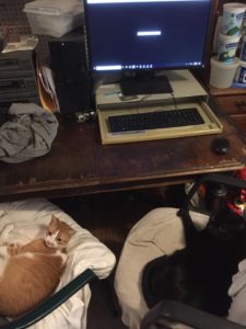
It’s a good thing that TWiV is a podcast.
Little Blake wanted to sit next to me by the computer, so I put a chair with a pillow for her there. Francesco must have thought that she looked comfy and then took my chair. It’s a good thing that with TWiV computer learning doesn’t mean being right at the computer.
Neva writes:
Hi TWIV-issimos,
This article was fascinating to me. It highlights not only a little known virus and touches on many aspects of the difficulty of addressing these things.
Thought this might be an interesting snippet for you.
Keep up the fabulous podcasts,
Neva of Buda
‘People are scared’: the fight against a deadly virus no one has heard of
Lucian writes:
Hi TWiVsters,
Thank you very much for the recent discussion on the decline in efficacy of the mumps vaccine. I took Michael Emerman’s virology course at the University of Washington last year and, for a final project, put together a presentation on mumps, and thought I might have some extra information to add:
The CDC did look at the use of a 3rd antibody, boost, and the results, though not terrible, were not particularly stellar. Antibody titers in individuals who received a 3rd booster were “highly correlated with baseline titer”; that is, the effect was minimal. During outbreaks where a 3rd MMR dose was tried, disease incidence did go down, but the 3rd dose was not administered until after the peak of the outbreak and it is unclear if the decline was affected by the booster or if it was the natural end of the outbreak. (Latner, Hickman, “Remembering Mumps,” PLoS Pathogens, 2015, 11(5); Marin, “Update on Mumps Epidemiology in the United States, 2017 and Review of Studies of 3rd Dose of MMR Vaccine,” CDC)
Part of what makes designing or evaluating vaccine strategies for mumps difficult is that there are no correlates of protection. And, while wild-type infection does seem to confer better immunity, titers from wild-type infection often aren’t all that great, either. Yoshida, et al., report that wild-type reinfection can occur in individuals—they even found patients who had been vaccinated and still had multiple wild-type infections. (Yoshida, et al., “Mumps virus reinfection is not a rare event confirmed by reverse transcription loop-mediated isothermal amplification,” J Med Virol, 2008, 80(3)).
One thought, which I think was mentioned in your discussion, is that it is possible that natural “boosting” occurred in vaccinated populations as a result of asymptomatic infections and, as vaccination has been successful, this boosting has gone away, allowing for new outbreaks.
Thank you for all the work you do!
Best,
Lucian DiPeso
Roton in the Moltke Lab
Molecular and Cell Biology Program
University of Washington
