Anthony writes:
Cosa Nostra in Hoboken
Bat vertical transmission:
Experimental Hendra Virus Infection in Pregnant Guinea-pigs and Fruit Bats(Pteropus poliocephalus)
https://www.sciencedirect.com/science/article/pii/S002199759990364X
fetuses are examined and vertical transmission is observed.
Here
Amplification of Emerging Viruses in a Bat Colony
https://www.ncbi.nlm.nih.gov/pmc/articles/PMC3165994/
bat droppings collected from below the colony are analysed for quantities of virus and vertical transmission is an interpretation. Bats generally go into a daytime torpor. Mother bats maintain a high metabolic rate during the day. I wonder if the the increase in virus shed is from adults. And for/if the juveniles that become infected, there’s no way to know from this paper if the transmission is infection from the mother before birth, during birth, from milk or by a vector (e.g. mite), from the air, etc.
Effect of rabies on bats:
This study seems to show that bats do die from rabies.
http://www.ncbi.nlm.nih.gov/pubmed/15966107
The common advice to avoid bats acting strangely is good to follow. The recent fatality in FLA was a child bitten by a visibly sick bat that the father was trying to rescue.
Islam writes:
Dear Vincent,
While responding to a listener’s email in TWiV 484, which was asking about influenza in seals, you asked whether there are any active surveillance programs.
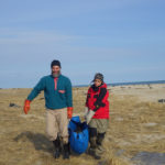
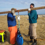
My ex-research group, headed by Professor Jonathan Runstadler and currently located at Tufts Veterinary School, is actually doing that. I was extremely lucky to have been associated with this excellent lab for few years, during which I learned a lot about field work of wild influenza. Since 2013, Professor Runstadler and his group has been going out every January to the south shore of New England to sample seals. They consistently find serological evidence of influenza infection. However, isolation and subtyping of these viruses has been challenging. In a recent paper published in Emerging Microbes and Infections, they proposed that the North Atlantic gray seals could serve as wild influenza reservoirs: https://www.nature.com/articles/emi201677
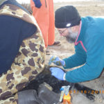
Here is an aerial picture of the team while sampling seals in the islands of Muskeget and Monomoy in January 2016: https://runstadlerlab.mit.edu/news/runstadler-lab-continues-sampling-seals-influenza-muskeget-and-monomoy-islands
These influenza disease ecology research efforts are funded through NIAID’s Centers of Excellence of Influenza Research and Surveillance, aided by NOAA’s Northeast Fisheries Science Center and other collaborators. The capture and sampling techniques are quite clever. Here is a glimpse in this press release by NOAA: https://www.nefsc.noaa.gov/press_release/pr2015/scispot/ss1501/
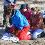
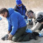
I am also attaching few pictures that demonstrate the extreme care with which these cute animals are captured, sampled and released peacefully. I think you might like to hear more about these amazing field adventures and all the tough logistics that go behind them from current members of the Runstadler lab, in particular my friends Dr Wendy Puryear and Dr Nichola Hill. Therefore, I am proposing a TWiV about wild influenza from Tufts Vet School during your next visit to MA and I am willing to coordinate the whole thing.
More about this interesting research can be found in this nice profile by the Tufts magazine: https://tuftsmagazine.com/issues/magazine/flu-hunters
And a recent blog post by Professor Runstadler in the Conversation: https://theconversation.com/influenzas-wild-origins-in-the-animals-around-us-91058
If the listener is interested in learning more about influenza in marine mammals, I suggest these two excellent reviews by Fereidouni et al., published in 2016 in EcoHealth: https://link.springer.com/article/10.1007%2Fs10393-014-0968-1
And by Virginia White published in 2013 in the US Army Med. Dep. J.: https://www.ncbi.nlm.nih.gov/pubmed/23277445
I am attaching PDFs of both.
By the way, what is the plan for TWiV 500? 🙂
Best,
Islam
Hi TWiV team,
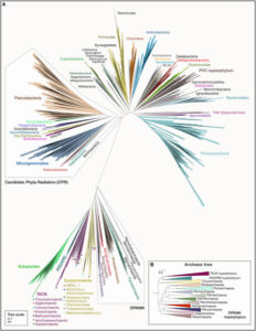
I just had a quick comment relating to Kathy’s episode 484 pick about the different formats of phylogenetic trees, so this might stray dangerously close to TWiEVO territory – I hope your listeners don’t mind! I admit I share her general distaste for irregular ways of presenting trees intended to actually be interpreted for meaning rather than for purely artistic purposes. However, I listened to the episode in the afternoon, and that morning I read a brand-new perspective paper that reminded me of the occasional utility of these layouts. Specifically, the radial format.
The paper is “Major New Microbial Groups Expand Diversity and Alter our Understanding of the Tree of Life” by Castelle and Banfield, published in Cell on March 8th.
http://www.cell.com/cell/fulltext/S0092-8674(18)30160-0
Their figure 1A (copied below) is a beautiful radial tree that in my opinion perfectly illustrates the 2-domain hypothesis of life. In contrast to the traditional “3-domain” tree of life where Eukaryotes, Bacteria, and Archaea are consider the major domains, more recent discoveries of groups of Archaea lead to a phylogenetic tree where Eukaryotes are firmly couched within the Archaea. In other words, “Archaea” are now considered paraphyletic rather than monophyletic. I don’t think a more traditional phylogenetic tree would illustrate this nearly as well, especially things like the distance between the Archaea+Eukaryote domain and the Bacteria domain, so in this instance I personally think the radial tree was a great choice.
To be honest I just really liked this tree so wanted you all to see it too!
Keep up the great work on the TWiX podcasts!
All the best,
Jake Leyhr
PS – The weather here in Uppsala, Sweden is a pleasant -2C with blue and sunny skies. It looks like we’re finally coming to the end of the long winter, although the snow isn’t going anywhere just yet.
Daniel writes:
Dear TWiV-Team,
this recent paper about functional insulin-like peptides produced by some Iridoviridae might pique your interest (I wonder what the exact function of these peptides are in the viral life cycle):
http://www.pnas.org/content/early/2018/02/20/1721117115
Keep up the great work!
Regards,
Daniel
————————————————————————————-
PhD Student, ETH Zürich
Meg writes:
Hello TWiV Posse!
Greetings from MoCo in the DC burbs, where it’s too windy to matter what the temperature is outside. My cell phone says 42F, 6C. Stuff is flying around out there! They closed NIH today so I got a chance to catch up on my reading. Roofers are going to be needed around here tomorrow from what I saw while walking my dog and dodging debris. I don’t know how many card-carrying neuroscientists you have among your listeners, but you have at least one. Like many scientists, I’m an artist at heart with a day job. You can see my brain art on Twitter @BrainsRus. I have an interest in virology and use viruses in my research. We use AAV mostly to carry fluorescent proteins or light-activated ion channels for “optogenetics” and neurophysiology experiments. I am a neuroanatomist and neuropharmacologist but I work closely with electrophysiologists and people who study behavior. I included my lab information below, which explains it all, except how I came to study cannabinoids. I’m still trying to figure that out!
I think you all did a great job with the recent Arc neuro papers. I’m also fascinated by this work so I’m glad you covered it. This brings me to the reason for this email that might involve a similar mechanism. It’s at least an interesting paper. Alan suggested that I write. There’s a problem with viral tract tracing that I keep encountering while others close their eyes or don’t realize what they are seeing. This recent paper by Zinng and colleagues confirms my observation. “AAV-Mediated Anterograde Transsynaptic Tagging: Mapping Corticocollicular Input-Defined Neural Pathways for Defense Behaviors” DOI: https://doi.org/10.1016/j.neuron.2016.11.045. Open Access. They use this to their advantage, which is great. The bigger picture could be a problem though. The supporting data were more informative for me. An injection into certain regions of cortex will infect cells in downstream areas in striatum in my hands too. This could be retrograde transport, from the axon terminals to the cell bodies, but I see it in pathways that are unidirectional like cortex to spiny striatal projection neurons, so it must be anterograde. Scratch that idea.
The second possibility is that there is transsynaptic transport of AAV, as this paper suggests. If this happens, how did the virus travel down a tiny sub-micron sized axon full of microtubules, and all of the things a neuron needs to actively transport to the synapse, and emerge as an infectious virus? Even rabies can’t do that, right? Doesn’t it need to replicate to cross a synapse? The AAVs are replication defective so it must be the original input. Another idea is as old as tract tracing itself. The cells died and released the tracer, but the virus in a capsid, it still has to get to the axon terminal. The next question is what is actually released from the virus-infected cell synapse. If it’s intact virus, that means there was transport down axons full of microtubules and packed like cables for 5 mm or so. Brain fiber tracts aren’t like blood vessels, so transport must have happened inside the cell. AAV is small so maybe it hitchhiked a ride on the transport machinery and was released at (possibly dying) synapses. Or maybe an Arc virus-like particle. There has to be a lot of it though. Not sold on this idea.
Could it be DNA or protein released? The recent Arc papers made me wonder if Cre is packaged and released, but this can’t be the whole story. It would need to be something that’s amplified, like Cre mediated expression of a fluorescent protein in the post-synaptic cell in this paper, in order to see it. Maybe it happens more than we realize but the virus never makes enough fluorescent protein to be detected? PCR from a distal target would tell us. I disagree with their conclusion with the GFP virus because the worst offender in my hands is the general AAV9 CaMK promoter driving mCherry. It has to be intact virus or DNA, and a lot of it, or we would never see the fluorescent protein from a CaMK promoter in the post synaptic cell. It could be naked DNA and just a function of the amount of virus people use. Our viruses come titered based on particles, not infectivity, but are usually in the neighborhood of 1013 particles/uL. We typically inject 100 nL into brains. That’s a lot of virus- national debt range into a tiny mouse prefrontal cortex!
This brings me to another even crazier idea. This is actually my favorite hypothesis. The AAV DNA was packaged into something else. Say the mouse has a virus that is similar enough to the AAV packaging requirements to package the replication defective AAV genome into a different capsid- like the old AAV in a herpes capsid strategy but with a mouse herpes-like virus. I think you only need two factors, rep and cap, supplied in trans to package AAV, right? What are the odds of co-infection?
We are getting ready to use the AAV1-Cre strategy from this paper. It would be interesting to hear an opinion from “actual virologists”. How is this happening? Did I cover all of the possibilities? This would be fun to try to figure out in the lab but we’re not equipped. AAV is supposed to stay put and this is too big of an artifact to just ignore. You can’t have infected local cell bodies when you’re trying to stimulate axons. Those cells have local collateral axons going to the neighbors too. See the problem? It’s great for me though since I’m an anatomist. I get to see who’s hooked up to who without resorting to rabies-based strategies.
I hope the TWIV brain collective will consider my questions and this paper for a TWiV discussion!
Thanks,
Meg
Margaret I. Davis, PhD
Staff Scientist
Section on Synaptic Pharmacology
Laboratory for Integrative Neuroscience
National Institute on Alcohol Abuse and Alcoholism
National Institutes of Health
Wink writes:
Dear TWIV Professors:
Why are tenofovir and lamivudine effective in the control of two very different viruses: hepatitis B and HIV?
Wink Weinberg (Atlanta)
Doug writes:
Dear wise sages of TWIV,
I am a software developer and white hat hacker with a speciality in medical informatics. I listen to TWiX podcasts when I can and love Dr. Vincent Racaniello’s youtube video course on virology. The parallels of virology and computer viruses are astonishing. I loved “TWiM 125<https://www.microbe.tv/twim/twim-125/>: A minimal cell operating system” as the TWiMsters discussed the J. Craig Venter Institute’s efforts to find the minimal bacterial genome.
One of the best ways to learn about computers is to hack on them and learn by trial and error. It seems like biohacking holds the same promises. I wonder how you feel about this recent youtube video: Developing a Permanent Treatment for Lactose Intolerance Using Gene Therapy (https://www.youtube.com/watch?v=J3FcbFqSoQY).
As a software developer this is exciting, but as someone who does not understand the risks in the field of virology, it’s hard for me to know how to feel about this self experimentation. I feel that biohacking on humans should be allowed, especially if the hacking is on rare and/or terminal diseases, but it’s hard to gauge based on the risks many point out in the comments section of the video.
Would love to hear your learned debate.
Thanks,
–Doug.
PS: I missed the chance to see TWiV 466/TWiM 164 live when Dr. Racaniello was at IU. Is there a schedule of upcoming visits to other universities?
—-
Douglas R. Horner
Medical Informatics Engineering, Inc. http://mieweb.com
6302 Constitution Dr., Fort Wayne, IN 46804
Paul writes:
Dear Twivians
Thanks for all your ongoing and enjoyable discussions on everything viral – just finished listening to last weeks podcast on the STING in a bat’s interferon response. Muggy here in Brisbane with the threat of a cyclone warning hanging over us!
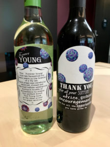
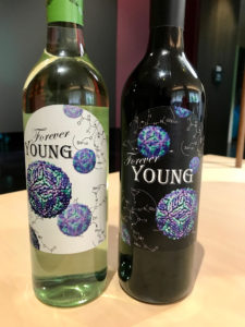
I thought I would share some pics of a going away present one of my recently graduated PhD students just gave me. Ashleigh (Shannon) is heading off to Marseilles to work with Bruno Canard on her first post-doc, funded by an Endeavour Fellowship. I’m sure she will have a great time there – particularly with the fantastic seafood!
Ash’s PhD was looking at pathways to antiviral drug design targeting the WNV protease. The labels on the two bottles of wine captured the essence of her PhD beautifully. I particularly liked the description on the back of one describing the grape origins 🙂 The “Forever Young” reference on the front is a nod to the name my group adopted whenever we entered a team in a Departmental social competition.
This is what Science is all about. Wonderful colleagues and life long friendships forged in the pursuit of discovery.
Keep up the great work!
Paul
Prof Paul R Young
School of Chemistry & Molecular Biosciences
University of Queensland
Brisbane 4072
Australia

