
Case guesses:
Sara writes:
Hi all,
I sadly have zero time for a lengthy analysis of this week’s case so this will be short and sweet! I’ll roll the dice on Toxocara canis or Ascaris lumbricoides.
Fingers crossed, looking forward to the next episode as always!
Sara (currently in Sydney)
Ben writes:
Dear TriTWiPomonads,
Firstly I need to clear the air in regards to South Australian geography and the pronunciation of Adelaide, Dickson had it right when he said he pronounced it “Add-elle-aid” but said that us Adelaideans would pronounce it “Add-elle-eyed”. We do have somewhat strange accents here though, I usually get asked if I’m English before Australian when I travel. Adelaide is about 8 hours along the coast from Melbourne and it is not in the Grampians but you do have to go past the Grampians to get here. The city of Adelaide is flanked by the Adelaide Hills, Barossa, Clare and Eden Valleys, which makes it perfect location for wine lovers! On top of this, we are right by the beach. Where I live now I am 15 minutes away from the beach, wine country and my university so I’m definitely going to be sad to see this place go when I hopefully move for a post-doc in the future!
I tried to do my research on this case but I was really struggling. It was impossible to hear a child has passed a worm of that length and not start thinking of Ascaris straight away but then the child had taken albendazole and it was a few months later and it didn’t all sound right. So here are my options (1), the serpiginous objects are Ascaris lumbricoides. This seems a little on the small size from what I’ve read, but perhaps the worms only get to a smaller size in a child. The possibilities I see for HOW the child could be infected with Ascaris include albendazole resistance, child was infected with Ascaris before coming back to US but the worms were resistant to the drug (not sure if this is often observed). Albendazole may not be effective against Ascaris in some of the earliest stages of infection, I couldn’t find anything supporting or refuting this. The albendazole might have been taken sufficiently long enough before coming back to the US that the child contracted Ascaris between albendazole treatment and leaving. Apparently Ascaris worms can take longer than 3 months to be passed in rare cases so that could explain any of the three scenarios above. (2) The child has ingested something that is not normally considered a parasite but has managed to survive through the GI tract and grow into a worm. I think I remember a case a while ago of someone passing worms after eating mangoes. (3) The child has eaten one of the items on the extensive list of things that kids have eaten and the parasites only find the evidence in their nappies.
A little side note I think you will like is the cover for the latest edition of the International Journal for Parasitology (only full size image I could find is on their facebook page https://www.facebook.com/IJPara/). It is a parody of the Beatles album let it be, that they’ve dubbed “Let it Babesia” J
Also a suggestion for a paper https://www.nature.com/articles/s41586-019-1072-z (although sadly behind a paywall). Parasitic apicomplexan that commonly infects coral, yet retains chlorophyll biosynthesis genes, fascinating stuff!
Thanks again for Podcasting a parasitic spell over me J
Regards,
Ben
Ben Liffner
PhD Candidate – Malaria Biology Lab (Wilson Lab)
Research Centre for Infectious Diseases
School of Biological Sciences
The University of Adelaide
Nathan writes:
Hello TWiP Experts,
This answer to this week’s case should be obvious, but there is one piece that makes me question my thinking. I’ll start by listing the important details that Dr. Griffin mentioned.
1. 18-month-old child traveling abroad with family.
2. Has been back in the US for 2-3 months.
3. Took a single dose of albendazole before leaving.
4. Has been in frequent contact with soil.
4. 18-month-old passed a 5-6-inch roundworm at daycare.
Based upon the size and shape of the worm, it sounds like the child was infected with the giant intestinal roundworm – Ascaris lumbricoides. Dr. Despommier describes this organism as being the “size of a pencil” (7.5 inches).
This case is curious because a single dose of albendazole should kill adult worms and prevent eggs from hatching. Albendazole disrupts glucose uptake and depletes glycogen store in the worm.
There are a few things that I think could possibly be going on:
1. The child did not take the dose of albendazole, so the worms were not eliminated.
2. The child became infected AFTER taking the medication after returning to the United States.
3. It is possible that the organism is resistant to albendazole and the treatment did not work. There have been some recent studies that have questioned the efficacy of Albendazole against Ascaris lumbricoides: For example: Reduced efficacy of albendazole against Ascaris lumbricoides in Rwandan schoolchildren was published in the International Journal for Parasitology: Drugs and Drug Resistance (2017)
4. The worm isn’t a parasitic worm. Maybe a common earthworm found its way into the stool. (I can’t believe that I’m even bringing this up, but it was seen in one of the labs where I used to work).
It sounds like the family may have to find a new daycare to attend!
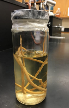
P.S. I have included an image of our preserved worms that we show our students. I have named them collectively: Elanor.
I also have a question for Dr. Despommier. When I took a parasitology course almost 10 years ago, the instructor mentioned that he traveled to Portugal with his family when he was young. He said that one of his extended family members was infected with Ascaris lumbricoides and that his family put a bowl of warm milk under his chin and the worms migrated out of his mouth.
I recently looked up using a bowl of milk as a treatment for Ascaris lumbricoides online and as I expected, there isn’t any information available. Was my instructor telling a tall tale? Have you ever heard a similar story?
Nathan
Lucian writes:
Dear TWiPsters!
The only parasite I can think of that would be 5 inches in length and round is Ascaris. As to why the child was infected despite treatment with albendazole, are there periods in the parasite’s life cycle where albendazole is less effective? I could find no mention of this, but it strikes me that perhaps while the parasite is in the lungs or perhaps while it is in the liver, concentrations of albendazole may not be sufficient to kill the parasite (in fact, I’m not sure you’d want the worm to die in your lungs!). Since maturation can take months, this could explain the delayed discovery of infection after treatment.
Many thanks for all you do!
Dr. Anderson’s Parasitology Class North Greenville University writes:
Dear TwIParasites:
It’s a stormy day in Tigerville, SC. Our parasitology professor is a big fan of your podcast. Below we have compiled a class response to the case study presented in the last episode.
We first considered identifying the parasite based on the exposure the patient had in a remote location outside of the US. Using the information provided by Dr. Griffin related to the size of the parasite, we narrowed it down to Ascaris lumbricoides and Wuchereria bancrofti.
Considering Ascaris:
- We know that this helminth is endemic in poverty stricken areas such as the one described in the case
- Further, considering the day to day activities of the child the embryonated egg was most likely ingested when the child was playing in the dirt
- The size of the organism the daycare worker encountered aligns with the average size of an adult male ascaris worm being 5.9-11.8”.
- The description of a roundworm rather than a flatworm also led us to this conclusion.
Considering Wuchereria:
- This worm is also a roundworm that can grow up to 5-6” and takes 3-6 months to develop into a mature adult stage.
- However, since this infection typically involves the lymphatic system we were able to eliminate it as a potential diagnosis.
Considering the unique presentation of the case with the delayed appearance of the worm, we have researched a few options to try to explain this outcome.
- First, low parasitic loads are usually asymptomatic as seen in the patient so the medication could have been partially effective
- We also are unsure if the dose provided to a child under 2 years old would be effective to eliminate the infection
- Typically ,as noted in a study by Adegnika et.al in 2014, one dose of albendazole is effective at clearing 85% of adult worms; however, we are curious if the treatment is not as effective at killing the migratory larval stages of the parasite.
- This would lead to an adult emerging 2- 3 months post treatment which may explain the organism encountered by the daycare worker
With the information provided in the case study, we don’t have a clear explanation (and are still thinking on) what would’ve caused the immediate evacuation of the worm—we considered that perhaps the child has developed a fever or some other illness that is causing the parasite to evacuate the premises.
We hope to win the book—it would be an excellent resource for our school!
Stay parasite free,
Dr. Anderson’s Parasitology Class North Greenville University, Tigerville SC
Heidi Dodson Anderson, MPH, Ph.D.
Assistant Professor of Biology
North Greenville University
Kevin writes:
Dear Twip professors
Not sure why my submission did not get received, but I’m sending it along so that perhaps it can be put into the show notes. Check out my elephantiasis anecdote under “A terminal curiosity”
K
TWiP 165 notes—Case read on TWiP 165 for discussion on TWiP 166
01/28/19
Kevin Carney
An adolescent male from eastern Uganda presents to Dr Griffin’s clinic with a painless scrotal mass. This approximately 6 centimeter swelling transilluminates when a light is placed behind the mass.
I begin with a quote from the English surgeon Percivall Pott (1714-1788), who many will remember elucidated the nature of “the soot ulcer”, scrotal carcinoma of chimney sweeps, one of the first occupational illnesses to be clearly described. Pott also described tuberculosis spondylitis and developed many surgical techniques, among those being correction of hydrocele: “An hydrocele is so irksome a disease to the indigent and laborious, furnishes even the easy and opulent with such disagreeable ideas and apprehensions, and is to all who are afflicted with it so troublesome and inconvenient, that every rational attempt toward relieving mankind form such an evil, will, I make no doubt, be favourably received.”
The difficulty in this case is not so much making the diagnosis, but rather grasping the enormity and complexity of the overall problem. The WHO estimates that almost 40 million persons are afflicted with some manifestation of lymphatic filariasis, with at least 25 million men suffering with hydrocele and 15 million persons with some degree of lymphedema. The offending agent in this case, and the culprit in over 80% of global parasitic lymphedema morbidity is Wuchereria bancrofti. In order not to fall into the habit of “System 1 thinking” however, a modest differential should be constructed. Other causes of scrotal swelling to consider: tuberculosis, cancer and indirect inguinal hernia. The fact that the mass transilluminates and the presence of many similar cases in the region makes W. bancrofti infection the overwhelming favorite. Chukwudi (2011) supports the use of transillumination as the sole diagnostic test in typical cases, and recommends forgoing the expensive and often unavailable scrotal ultrasound. Other diagnostic modalities: peripheral blood smear for microfilaria as well as field-ready immunologic tests such as immunochromatographic cards. In summary, typical presentation, regional prevalence and absence of other complications makes the use of “clinical diagnosis” -i.e. using you mind and hands – sufficient for definitive diagnosis in our case. Treatment of this condition poses some dilemmas. Conventional antihelminthic treatment (diethylcarbamazine) is a fairly poor macrofilaricidal agent and may even worsen the lymphatic pathology. Newer antibiotic approaches using doxycycline are being used with published data describing improvement in hydrocele pathology and progression. Doxycycline targets the endosymbiont Wolbachia which is believed to be crucial in the immunopathogenesis of lymphatic filariasis and hydrocele, via a variety of mechanisms involving VGEF, TNF, interleukins etc. Surgical management (using conventional surgery or sclerotherapy) is undertaken in selected cases but is often unavailable. Below references discuss patient selection etc for surgery. In addition to pharmacologic and surgical management, treatment must also include simple modalities such as scrupulous hygiene of the involved genitalia, since concomitant bacterial and fungal infection as well as generalized skin inflammation and fibrosis can contribute to the progression of the disease. It must be emphasized that tropical hydrocele is a cause of considerable psychological, social, sexual, and employment disability and mitigation of these factors must be included in the care of the patient. Further reading and background information is included in the endnotes, as well as near irrelevancies such as podoconiosis and a further examination of disease metaphors, this time of a zoological nature…(see culinary metaphors digression- the anchovy sauce affair in TWiP 159 case notes-Toddy Tappers). Speaking of medical terminology, please do not overlook the rather non-technical definition of ‘giant hydrocele’ in the Akpo reference below.
Thanks for your ongoing stimuli, which has revived my cortex after the polar vortex.
END NOTES AND A TERMINAL CURIOSITY
General Filariasis/ W bancrofti:
TWiP 25: Wuchereria bancrofti April 27, 2011, https://www.microbe.tv/twip/25-wuchereria-bancrofti/
A 2015 eight minute video from the BBC world service on lymphatic filariasis in India, primarily focussed on the mass drug administration program and its “operational challenges” : https://www.bbc.co.uk/sounds/play/p02jcqnp
This brief video is well worth viewing.
Manual of Tropical Medicine, (1242 pages) Sir Aldo Castellani, Albert John Chambers, William Wood & Company, 1910. Massive treatise available free via books.google.com. Interesting chapter on the filariases. Nice companion to PD6. No messy immunology or cellular biology.
Current Epidemiological Assessment of Bancroftian Filariasis in Tanga Region, Northeastern Tanzania
Happyness J. Mshana et al, J Trop Med. 2016; 2016: 7408187. OPEN ACCESS
Even after mass drug administration begun in 2000 for filiariasis in Tanzania, n=472, 5% had circulating filarial antigen, prevalence of hydrocele was 73%, lymphedema=16% Our findings demonstrate a considerable reduction in filarial infection.
Guideline: Alternative mass drug administration regimens to eliminate lymphatic filariasis. Geneva: World Health Organization; 2017. Licence: CC BY-NC-SA 3.0 IGO.
quote from Pfarr: Mass drug administration (MDA) programs aim to interrupt mosquito vector transmission and eventually eradicate the disease. However, MDA does not help those who suffer from lymphedema or hydrocele, estimated to affect 12% and 25 % of infected individuals.
Filariasis and lymphoedema, Pfarr KM et al, Parasite Immunol. 2009 Nov;31(11):664-72. doi: 10.1111/j.1365-3024.2009.01133.x. OPEN ACCESS
Abstract: Among the causes of lymphoedema (LE), secondary LE due to filariasis is the most prevalent. It affects only a minority of the 120 million people infected with the causative organisms of lymphatic filariasis (LF), Wuchereria bancrofti and Brugia malayi/timori, but is clustered in families, indicating a genetic basis for development of this pathology…. Importantly, as for the aberrant lymph vessel development, innate immune responses that are triggered by the filarial antigen ultimately result in the activation of vascular endothelial growth factors (VEGF), thus promoting lymph vessel hyperplasia as a first step to lymphoedema development. Wolbachia endosymbionts are major inducers of these responses in vitro, and their depletion by doxycycline in LF patients reduces plasma VEGF and soluble VEGF-receptor-3 levels to those seen in endemic normals preceding pathology improvement. The search for the immunogenetic basis for LE could lead to the identification of risk factors and thus, to prevention; and has so far led to the identification of single-nucleotide polymorphisms (SNP) with potential functional relevance to VEGF, cytokine and toll-like receptor (TLR) genes. Hydrocele, a pathology with some similarity to LE in which both lymph vessel dilation and lymph extravasation are shared sequelae, has been found to be strongly associated with a VEGF-A SNP known for upregulation of this (lymph-)angiogenesis factor.
Wolbachia filarial interactions, Mark J. Taylor et al, Cellular Microbiology (2013) 15(4), 520–526
OPEN ACCESS
A very technical, cell biology oriented review. Taylor is funded by the Bill & Melinda Gates Foundation. The website of the anti-wolbachia consortium: https://awol.lstmed.ac.uk/
I find it extraordinary that a parasite is dependent on an obligate intracellular bacterium for development, oogenesis, embryogenesis, larval development and establishment, and as “an essential partner to key biological processes in the life of the nematode.”
Chronic clinical manifestations related to Wuchereria bancrofti infection in a highly endemic area in Kenya
S.M. Njenga et al, Transactions of the Royal Society of Tropical Medicine and Hygiene (2007) 101, 439—444
Malindi district, Kenya. Of 186 males aged 15 years and above examined, 64 individuals (34.4%) had hydrocele, and the prevalence of the manifestation in those above 40 years old was 55.3%. The prevalence of leg lymphoedema in persons aged 15 years and above was 8.5%, with a higher rate in males (12.6%) than in females (5.7%). The overall prevalence of inguinal adenopathy was 8.6%, and males had a significantly higher (12.9%) prevalence of adenopathy than females (5.1%) (P < 0.001)……Hydrocele was the most common chronic clinical manifestation of lymphatic filariasis observed in the present study….This observation is consistent with reports from most studies in sub-Saharan Africa.
Excellent public health advertisement/ consciousness raising mass media piece:
Two minute public health video from the Sabin Vaccine Institute and Richard Hatzfeld directed to affected communities in India to assist with raising awareness and enhancing mass drug administration adherence. Quite a touching advertisement.
https://www.campaignindia.in/video/taking-giant-steps-to-end-filaria-with-awareness-campaign/421987
EXCELLENT fact sheet on lymphatic filariasis from the WHO:
https://www.who.int/en/news-room/fact-sheets/detail/lymphatic-filariasis
Hydrocele:
Scrotal anatomy and the formation of hydrocele.
The testes are “suspended” in a potential cavity (the cavum vaginale) that is defined by two serous layers of the tunica vaginalis (the outer lamina parietalis and the inner lamina visceralis). It is this space that becomes distended by fluid that defines the clinical entity of hydrocele. ….blockage of lymphatic flow and chronic inflammation is believed to result in fluid secretion by the tunica layers with resultant swelling (hydrocele)
There is even an analog of hydrocele in women, Hydrocele of the Canal of Nuck (usually a congenital defect)
The Chirurgical Works of Percivall Pott FRS Vol III, 1779 (Free download at google.books.com)
Link below contains a very thorough review of filarial hydrocele, fairly nontechnical. Good illustrations.:
https://emedicine.medscape.com/article/438525-overview#a10
Reduction in levels of plasma vascular endothelial growth factor-A and improvement in hydrocele patients by targeting endosymbiotic Wolbachia sp. in Wuchereria bancrofti with doxycycline, Debrah AY, et al. Am J Trop Med Hyg 2009; 80: 956–963.
Discusses the effect of doxycycline on hydrocele…some emerging evidence for genetic / familial susceptibility to hydrocele development….people with SNPs that are associated with upregulated VEGF-A
WHO: The global baseline estimate of persons affected by lymphatic filariasis is 25 million men with hydrocele and over 15 million people with lymphoedema. At least 36 million persons remain with these chronic disease manifestations [https://www.who.int/lymphatic_filariasis/resources/9789241550161/en/]
Filarial hydrocele: a neglected condition of a neglected tropical disease, Kenneth Bentum Otabil, Seth Boateng Tenkorang, J Infect Dev Ctries 2015; 9(5):456 – 462 OPEN ACCESS.
A concise 7 page review. Filarial hydrocele is the MOST COMMON clinical manifestation of lymphatic filariasis.
Longstanding hydrocele in adult Black Africans: Is preoperative scrotal ultrasound justified?
Chukwudi O. Okorie, Nigerian Medical Journal. 2011 Jul-Sep; 52(3): 173–176.
Scrotal ultrasound is unnecessary in most cases. Transillumination is recommended. This study was a series of 102 patients, 97% were simple hydrocele, 3% had loculated hydrocele. hydrocele is more common on the right ( findings in 102 patients: left: 23 patients, right: 39, bilateral: 40.
An historical perspective:
HYDROCELE AMONGST THE LANGO OF UGANDA. WILLIAM P. KELLY, F.R.C.S.I., D.P.H.,
MEDICAL OFFICER (TEMPORARY), UGANDA MEDICAL SERVICE. The British Medical Journal, April 26, 1924
a description of a surgical technique to ameliorate this condition…interestingly, no mention of filiariasis is mentioned in the article. “THE natives of Lango (Uganda) do not appear to be possessed of any surgical skill whatever, and so pathological conditions amongst them tend to become very gross indeed. Hydroceles and, as a consequence, haematoceles are very common; some of these tumours grow to enormous proportions, often reaching nearly to the knee, causing great disability. The scrotal tissues are, as a rule, greatly thickened; and the disappearance.”
A Comparative Study of Sclerotherapy With Phenol Versus Surgical Treatment For Hydrocoele, Labib M A et al, East and Central African Journal of Surgery, Vol. 9, No. 2, Dec, 2004, pp. 25-27
Prospective trial of 80 consecutive hydrocele patients. The authors conclude: “Sclerotherapy for hydrocoele using phenol is as efficient as hydrocelectomy for cure. The risk of complications arising from phenol sclerotherapy is slight, while it allows the patient to return to normal activity on the same day, so sclerotherapy may be the option of choice for hydrocoele.”
Giant hydrocele – an epitome of neglect Emmanuel E Akpo, Afr Health Sci. 2005 Dec; 5(4): 343–344.
OPEN ACCESS. A case series. Akpo provides his own unique definition of giant hydrocele which for “clinical purposes, (is) a hydrocele equal to or bigger than the patient’s head.” Case 1, a 50 y/o man with bilateral scrotal enlargement to the knees and a ‘buried phallus’ had, upon surgery, 4 litres of fluid drained. Article contains some alarming photographs of the patient’s initial presentation.
Classifying Hydroceles of the Pelvis and Groin: An Overview of Etiology, Secondary Complications, Evaluation, and Management, Dagur G.Curr Urol. 2017 Apr;10(1):1-14
OPEN ACCESS. A massive review of ‘all things hydrocele’ …Fantastic diagrams of the many varieties of hydrocele. Did you know that there was a female variant of hydrocele?–you can read about the “Hydrocele of the canal of Nuck, also known as female hydrocele or cyst of the canal of nuck, affects infant females and results in painless or possibility painful inguinal swelling. It is an uncommon disease caused by the failure of the processus vaginalis to close during embryological development which can lead to inguinal hernia and hydrocele.” Management options discuss aspiration and sclerotherapy versus hydrocelectomy.
PODOCONIOSIS:
I was fascinated to learn about this condition, which is new to me. Wikipedia’s definition: “Podoconiosis, also known as nonfilarial elephantiasis, is a disease of the lymphatic vessels of the lower extremities that is caused by chronic exposure to irritant soils.” The condition is found in highland tropical Africa, northwest India and Central America. It is mentioned since it is peripherally related to our case in the context of lymphedema. Podoconiosis is seen in Uganda, although at low levels. It typically causes leg lymphedema and does not usually involve the scrotum or result in hydrocele. As if poverty, neglect and an array of insect vectors and parasitic worms were not enough, even the soil beneath the feet becomes a threat to health.
How Soil Scientists Help Combat Podoconiosis, A Neglected Tropical Disease, Benjamin Jelle Visser, Int J Environ Res Public Health. 2014 May; 11(5): 5133–5136. OPEN ACCESS
Global epidemiology of podoconiosis: A systematic review, Kebede Deribe, et al, PLoS Negl Trop Dis. 2018 Mar; 12(3) OPEN ACCESS
Non-filarial elephantiasis in the Mt. Elgon area (Kapchorwa District) of Uganda. Onapa AW, et al, Acta Trop. 2001 Feb 23;78(2):171-6.
Abstract
Elephantiasis was observed in all age groups from 10 years and above. The overall prevalence was 4.5%, and the prevalence among individuals aged >/=20 years was 8.2%. Males and females were equally affected. However, there were only few cases of hydrocele (overall prevalence in males of 1.0%) and blood examinations were negative for W. bancrofti circulating antigens and microfilariae.\In view of the low hydrocele to elephantiasis ratio, the absence of filarial infection in humans and mosquitoes, the high altitude (1500-2200 m above sea level) and the volcanic soil type, it is concluded that elephantiasis seen in this area is not of filarial origin but most likely is due to podoconiosis (endemic non-filarial elephantiasis).
A Terminal Curiosity:
Several years ago I visited a bedbound woman who weighed well over 500 pounds. In addition to the burdens of obesity she was afflicted with massive bilateral lymphedema of the legs. This American woman had never travelled outside of the US. She had a condition known as Elephantiasis nostras verrucosa. This entity is reviewed by Castellani- a famous tropical medicine researcher who authored the 1913 ‘Note on Copra Itch’ (referenced in TWiP 157). This is the only time I had encountered anything like the appalling photographs seen in tropical medicine textbooks.
During our visit I mentioned that her condition was known as elephantiasis nostras. The patient immediately became very distressed by the association of her lamentable state with elephants. It took my best medical casuistry to backpedal out of this morass and the whole episode taught me the profound subjectivity and emotional content of medical language as interpreted by patients.
The WHO published guidelines concerning the naming of diseases in 2015. (see refs below). The press predictably branded the WHO initiative as a species of political correctness and reported the story in a slightly snide or frivolous way. As the above anecdote shows, a name can have profound emotional impact. Naming diseases after geographic locations can even have economic consequences.
This topic stimulated me to recall the many medical terms that are named after animals, many of which would probably be very distressing to patients. Fortunately many of these terms are being replaced by more accurate descriptors.
List of zoological medical metaphors:
rodent ulcer basal cell carcinoma is a bit more neutral
maus kopf used to describe facial appearance in scleroderma
pigbel pidgen-english term for post-starvation gastroenteritis
lupus pernio lupus- the wolf, purportedly used to describe skin that appears ravaged as if chewed by a wolf
swine flu
bird flu
icthyosis who wants to be compared to a fish?
moth-eaten alopecia
monkey pox
mad-cow
leonine facies a classic term in lepromatous leprosy, Paget’s disease of bone and diffuse cutaneous leishmaniasis
buffalo hump just what your Cushingoid patients want to hear- additionally, if they hear cushingoid they probably think they are being compared with a cushion
butterfly rash not too bad as these things go
simian crease monkey analogies are to be universally avoided.
raccoon sign
phocomelia
bulimia
eqinovarus deformity
elephant-man syndrome
spider angioma
(for completness let us mention anatomic terms with animal associations: hippocampus, pes anserinus, vermiform appendix)
References to accompany ‘A Terminal Curiosity’:
Elephantiasis Nostras Verrucosa, Krisanne Sisto et al, Am J Clin Dermatol 2008; 9 (3): 141-146
Castellani A. Researches on elephantiasis nostras and elephantiasis tropica with regard to their initial stage of recurring lymphangitis (lymphangitis recurrens elephantogenica). J Trop Med Hyg 1969 Apr; 72 (89): 89-96
WHO issues best practices for naming new human infectious diseases
https://www.who.int/mediacentre/news/notes/2015/naming-new-diseases/en/
WHO best practices for naming of new human infectious diseases
https://www.who.int/topics/infectious_diseases/naming-new-diseases/en/
Don’t Name New Human Infectious Diseases After Animals Or Places, Says WHO: Here’s Why
Animals Eponyms in Dermatology, Nidhi Jindal, Indian J Dermatol. 2014 Nov-Dec; 59(6): 631. OPEN ACCESS
Favorite Animal Names in Dermatology, Walter H. C. Burgdorf, et al, JAMA Dermatology August 2013 Volume 149, Number 8
The term “elephantiasis” is more than 2000 years old and is mentioned by Celsus in his work De Medicina (Book III: 25). The term is derived from the Greek word for elephant, “elephas.”
The menagerie of neurology, Animal signs and the refinement of clinical acumen, Shin C. Beh et al, Neurol Clin Pract. 2014;4(3):e1-e9.
Anthony writes:
TWiP 162 – over half the animals are parasites.
There’s one species humans that hosts how many parasites? (hundreds?) Presumably, we’re not more infestation prone, just better studied than other animals. If that’s so, then it’s not just over half, but the vast majority of animals that are parasites.
FWIW
Anne writes:
Hello All,
A link on the importance of parasites…
https://www.theatlantic.com/magazine/archive/2019/03/iberian-lynx-parasite/580435/
Regards,
Anne
Anthony writes:
You’re doing a great job with science communication now, but perhaps through branding and a print medium MANY more might be reached: